Dive into the fascinating world of the cell cycle, the essential process governing cell growth and division across all living organisms. This comprehensive guide illuminates the stages from interphase to mitosis, highlighting the orchestrated events that enable cells to duplicate their DNA and split into two. Explore real-world examples that demonstrate the cell cycle’s critical role in development, tissue repair, and cancer research. Unlock the secrets of cellular replication and discover how understanding the cell cycle paves the way for breakthroughs in medical science.
What are Cell Cycle?
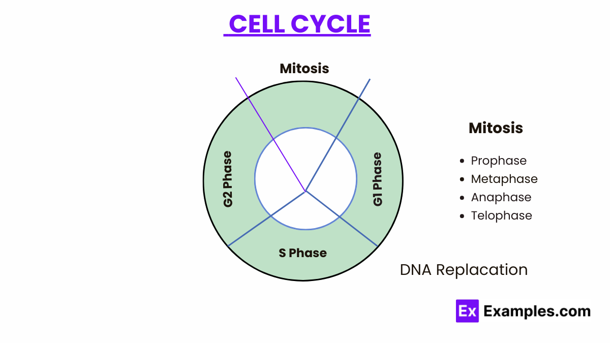
The cell cycle represents the series of events that take place in a cell leading to its division and duplication (replication). It is the cornerstone of cellular organization and growth, ensuring that cells reproduce accurately and healthy. The cell cycle is meticulously regulated and can be divided into two major phases: interphase and the mitotic (M) phase.
Phases of the Cell Cycle
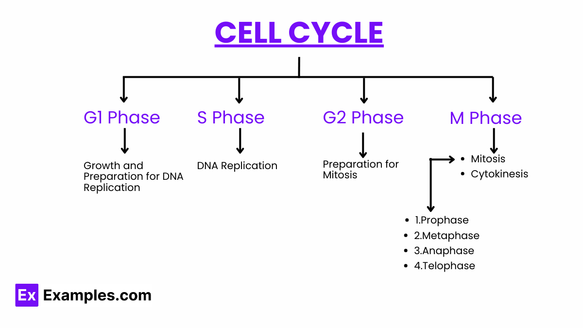
The cell cycle is divided into two major phases: interphase and the mitotic phase (M phase).
-
G1 phase (Gap 1)
The cell grows in size and synthesizes mRNA and proteins necessary for DNA replication. It’s a phase of metabolic activity and growth.
-
S phase (Synthesis)
DNA replication occurs, ensuring that each daughter cell will receive an exact copy of the cell’s DNA.
-
G2 phase (Gap 2)
The cell continues to grow and prepares for mitosis, synthesizing proteins and organelles needed for cell division.
-
Mitotic Phase (M Phase)
The mitotic phase includes mitosis and cytokinesis, leading to the physical division of the cell into two daughter cells
- Mitosis: Divided into four stages – prophase, metaphase, anaphase, and telophase – mitosis is the process through which the cell’s chromosomes are equally distributed between the two new nuclei.
- Cytokinesis: The cytoplasm divides, resulting in two separate daughter cells, each with a complete set of chromosomes.
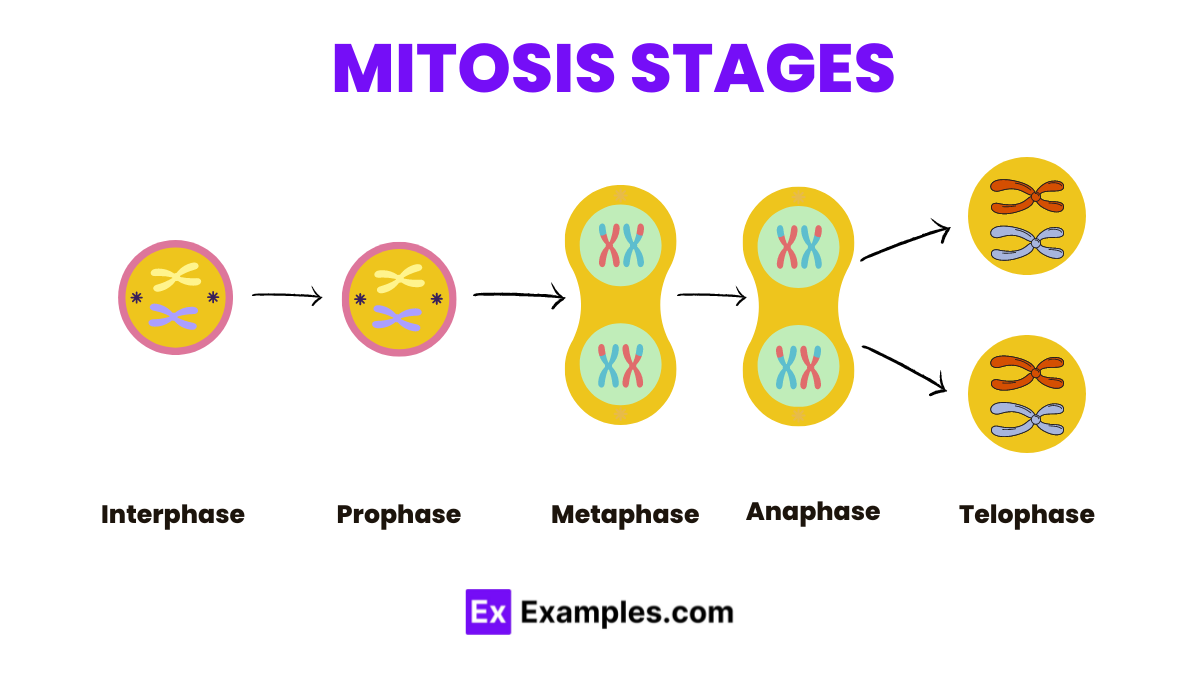
Mitosis Stages
-
- Interphase: Interphase is the period of growth and preparation for cell division.
- Prophase: Chromosomes condense, and the nuclear envelope breaks down.
- Metaphase: Chromosomes align at the cell’s equatorial plate.
- Anaphase: Sister chromatids separate and move to opposite poles.
- Telophase: Nuclear envelopes re-form around the separated chromosomes, which begin to decondense.
Regulation of the Cell Cycle
The cell cycle is tightly regulated by a complex network of signaling pathways, involving cyclins, cyclin-dependent kinases (CDKs), and various checkpoint proteins. These regulatory molecules ensure that each phase is completed accurately before the cell proceeds to the next stage, preventing errors such as DNA damage or incomplete replication that could lead to mutations or cell death.
Key Regulators of the Cell Cycle
The regulation of the eukaryotic cell cycle involves a sophisticated network of cyclins, cyclin-dependent kinases (Cdks), and various checkpoint proteins that ensure the cycle proceeds at the right time and in the correct order.
- Cyclins and Cyclin-Dependent Kinases (Cdks): Cyclins are proteins whose concentrations vary throughout the cell cycle. They activate Cdks, which are critical enzymes that phosphorylate specific target proteins to initiate or regulate cell cycle transitions. The cyclin-Cdk complexes are pivotal for the progression of the cell cycle phases.
- Cell Cycle Checkpoints: These are surveillance mechanisms that monitor and verify whether the processes at each phase of the cell cycle have been accurately completed before the cell progresses to the next phase. The main checkpoints are:
- G1 Checkpoint: Determines whether conditions are favorable for DNA replication.
- S Checkpoint: Ensures DNA has been replicated without errors.
- G2 Checkpoint: Checks for DNA damage post-replication and ensures all chromosomes are fully replicated.
- M Checkpoint (Spindle Assembly Checkpoint): Ensures chromosomes are properly aligned and attached to the spindle fibers before anaphase begins.
Regulatory Pathways
The eukaryotic cell cycle is also influenced by several signaling pathways that respond to internal and external cues, such as DNA damage, nutrient status, and cell size.
- DNA Damage Response: When DNA damage is detected, proteins like ATM and ATR activate p53, leading to cell cycle arrest until the damage is repaired. This prevents the propagation of damaged DNA.
- Growth Factors: External signals can stimulate cell proliferation. Growth factors bind to receptors on the cell surface, activating signaling cascades that culminate in cell cycle progression.
- Cell Size and Nutrient Availability: Eukaryotic cells also monitor their size and the availability of nutrients. Adequate cell size and nutrients are prerequisites for cell cycle progression, especially at the G1 checkpoint.
Therapeutic Implications and Research
Understanding the regulation of the eukaryotic cell cycle has profound implications for cancer therapy, as many cancers are characterized by deregulated cell cycle control. Targeting specific regulators of the cell cycle, such as cyclin-Cdk complexes or checkpoint kinases, offers a strategy for developing cancer therapeutics. Additionally, research into cell cycle regulation can provide insights into developmental biology, stem cell biology, and aging.
Fluorescence Imaging of the Cell Cycle
Fluorescence imaging of the cell cycle stands as a revolutionary technique in cellular biology, allowing researchers to visualize and track the dynamic processes of cell division in real-time. This method employs fluorescent markers to highlight specific proteins or structures within the cell, enabling the detailed observation of cell cycle progression, from DNA replication to mitosis. This comprehensive guide explores the principles, techniques, and applications of fluorescence imaging in studying the cell cycle, shedding light on its importance in advancing our understanding of cellular processes and disease mechanisms.
Principles of Fluorescence Imaging
Fluorescence imaging is based on the excitation of fluorescent dyes or proteins by specific wavelengths of light, causing them to emit light at different wavelengths. When these fluorescent markers are attached to molecules or structures of interest within the cell, they allow for the direct observation of cellular processes under a fluorescence microscope. The specificity and sensitivity of fluorescence imaging make it an indispensable tool for cell cycle studies.
Techniques for Cell Cycle Imaging
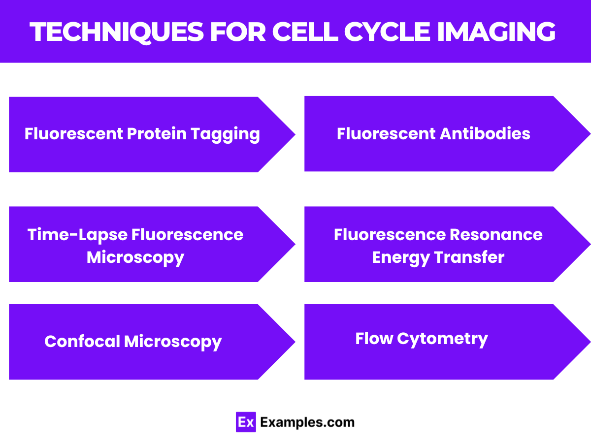 Several techniques are employed in fluorescence imaging of the cell cycle, each offering unique insights into the cellular processes:
- Fluorescent Protein Tagging: Genes encoding fluorescent proteins, such as GFP (green fluorescent protein), can be fused to genes of cell cycle regulatory proteins. This allows for the visualization of the location and concentration of these proteins throughout the cell cycle.
- Fluorescent Antibodies: Specific antibodies tagged with fluorescent dyes can bind to cell cycle markers, such as cyclins or phosphorylated proteins, highlighting their presence and distribution in the cell.
- FRET (Fluorescence Resonance Energy Transfer): FRET can be used to study interactions between cell cycle proteins by detecting energy transfer between two closely located fluorescent molecules, indicating protein-protein interactions.
- Time-Lapse Fluorescence Microscopy: This technique involves capturing a series of images over time, showing the dynamic changes in fluorescently labeled structures or proteins as the cell progresses through the cell cycle phases.
- Confocal Microscopy: Provides high-resolution images of cells by focusing a laser through a pinhole, allowing for the detailed observation of cell cycle events in three dimensions. It is particularly useful for studying the localization and dynamics of fluorescently labeled cell cycle proteins.
- Flow Cytometry: Analyzes the DNA content of cells stained with fluorescent dyes to determine their cell cycle phase based on the amount of DNA present. This technique is useful for quantifying the proportion of cells in different cell cycle stages within a population.
Applications in Research
Fluorescence imaging of the cell cycle has broad applications in biological research, including:
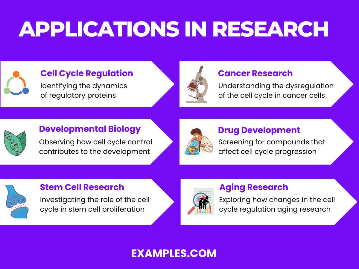
- Cell Cycle Regulation: Identifying the dynamics of regulatory proteins and their localization during different cell cycle stages.
- Cancer Research: Understanding the dysregulation of the cell cycle in cancer cells, helping to identify potential targets for therapy.
- Developmental Biology: Observing how cell cycle control contributes to the development and differentiation of tissues and organs.
- Drug Development: Screening for compounds that affect cell cycle progression, offering potential leads for anticancer drugs.
- Stem Cell Research: Investigating the role of the cell cycle in stem cell proliferation and differentiation, crucial for regenerative medicine and understanding tissue repair mechanisms.
- Aging Research: Exploring how changes in the cell cycle regulation contribute to the aging process and age-related diseases, providing insights into potential anti-aging strategies.
Challenges and Future Directions
While fluorescence imaging of the cell cycle provides invaluable insights, it also faces challenges such as phototoxicity, where prolonged exposure to light can damage cells, and photobleaching, where fluorescent markers lose their brightness over time. Advances in fluorescent proteins, imaging techniques, and computational analysis are continually being developed to overcome these challenges, promising even more detailed and accurate observations of the cell cycle in the future.
Cell Cycle Evolution
The evolution of the cell cycle is a fascinating journey through the history of life on Earth, reflecting the intricate changes that have enabled cells to proliferate, adapt, and evolve in response to environmental pressures and opportunities. This comprehensive guide explores the evolutionary advancements of the cell cycle, shedding light on how cellular replication mechanisms have diversified across different life forms and contributed to the complexity of life as we know it today. Understanding the evolution of the cell cycle not only illuminates the past but also provides insights into future directions in biology, medicine, and evolutionary research.
Origins of the Cell Cycle
The cell cycle’s evolutionary roots trace back to the earliest forms of life, with prokaryotic cells exhibiting simple division processes like binary fission. As life evolved, the need for more controlled and accurate cell division led to the development of the eukaryotic cell cycle, characterized by sophisticated checkpoints and regulatory mechanisms ensuring DNA integrity and proper segregation.
Comparative Cell Cycle Biology
Studying the cell cycle across different species has revealed both conserved mechanisms and unique adaptations. For example, the basic machinery of the cell cycle is remarkably conserved from yeast to humans, highlighting its fundamental importance. However, specific adaptations, such as those enabling certain plants and animals to undergo rapid cell cycles or dormancy periods, illustrate the cell cycle’s flexibility in response to ecological niches.
FAQS
What are the different phases of a cell cycle?
The cell cycle comprises distinct phases: G1 (cell growth), S (DNA synthesis), G2 (preparation for mitosis), and M phase (mitosis and cytokinesis), regulating cell division and replication.
What are the four major stages of mitosis?
The four major stages of mitosis are prophase, metaphase, anaphase, and telophase, which describe the process of chromosome condensation, alignment, separation, and division into two nuclei.
What do you understand by cell cycle?
The cell cycle is a series of regulated steps a cell undergoes leading to its division and duplication, ensuring accurate DNA replication, division, and cell function.
The cell cycle is a fundamental process governing cell growth and division, essential for life. Its intricate regulation ensures genetic fidelity and proper cellular function, impacting development, repair, and disease. Understanding the cell cycle’s mechanisms offers valuable insights into biology and medicine, providing a basis for innovations in cancer therapy, regenerative treatments, and genetic research, marking its significance in the science of life.