CheXNet:
Radiologist-Level
Pneumonia
Detection
on
Chest
X-Rays
with
Deep
Learning
Pranav
Rajpurkar
* 1
Jeremy
Irvin
* 1
Kaylie
Zhu
1
Brandon
Yang
1
Hershel
Mehta
1
Tony
Duan
1
Daisy
Ding
1
Aarti
Bagul
1
Robyn
L.
Ball
2
Curtis
Langlotz
3
Katie
Shpanskaya
3
Matthew
P.
Lungren
3
Andrew
Y.
Ng
1
Abstract
We
develop
an
algorithm
that
can
detect
pneumonia
from
chest
X-rays
at
a
level
ex-
ceeding
practicing
radiologists.
Our
algo-
rithm,
CheXNet,
is
a
121-layer
convolutional
neural network trained on ChestX-ray14, cur-
rently
the
largest
publicly
available
chest
X-
ray
dataset,
containing
over
100,000
frontal-
view
X-ray
images
with
14
diseases.
Four
practicing
academic
radiologists
annotate
a
test
set,
on
which
we
compare
the
perfor-
mance
of
CheXNet
to
that
of
radiologists.
We
find
that
CheXNet
exceeds
average
ra-
diologist
performance
on
the
F1
metric.
We
extend
CheXNet
to
detect
all
14
diseases
in
ChestX-ray14 and achieve state of the art re-
sults
on
all
14
diseases.
1.
Introduction
More than 1 million adults are hospitalized with pneu-
monia
and
around
50,000
die
from
the
disease
every
year
in
the
US
alone
(
CDC
,
2017
).
Chest
X-rays
are
currently the
best
available
method
for
diagnosing
pneumonia (
WHO
,
2001
), playing a crucial role in clin-
ical
care
(
Franquet
,
2001
)
and
epidemiological
studies
(
Cherian
et
al.
,
2005
).
However,
detecting
pneumo-
nia
in
chest
X-rays
is
a
challenging
task
that
relies
on
the availability of expert radiologists.
In this work, we
present
a
model
that
can
automatically
detect
pneu-
monia from chest X-rays at a level exceeding practicing
radiologists.
*
Equal
contribution
1
Stanford
University
Depart-
ment
of
Computer
Science
2
Stanford
University
De-
partment
of
Medicine
3
Stanford
University
Depart-
ment
of
Radiology.
Correspondence
to:
Pranav
Rajpurkar
<
pranavsr@cs.stanford.edu
>
,
Jeremy
Irvin
<
jirvin16@cs.stanford.edu
>
.
Project
website
at
https://stanfordmlgroup.
github.io/projects/chexnet
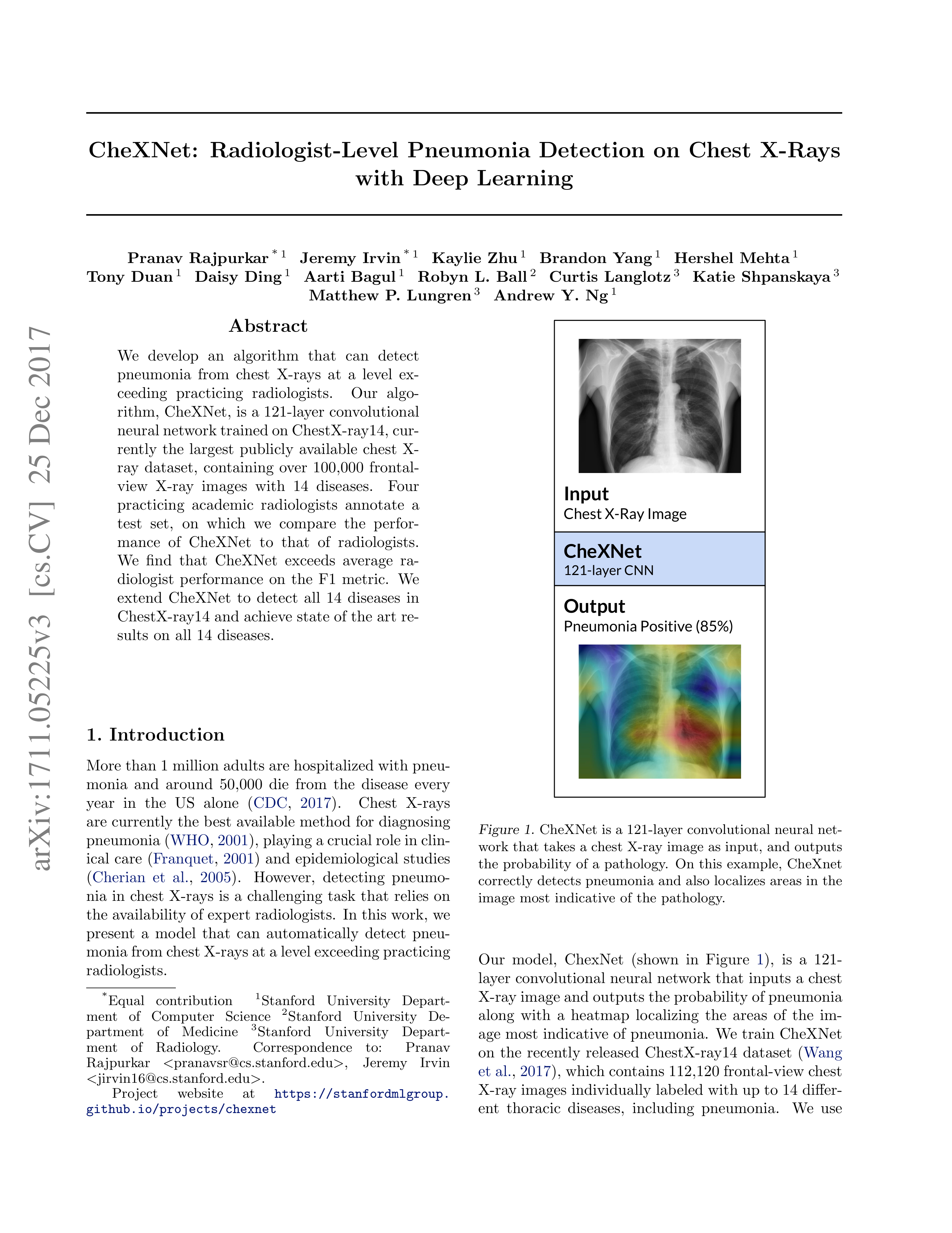
Output
Pneumonia Positive (85%)
Input
Chest X-Ray Image
CheXNet
121-layer
CNN
Figure
1.
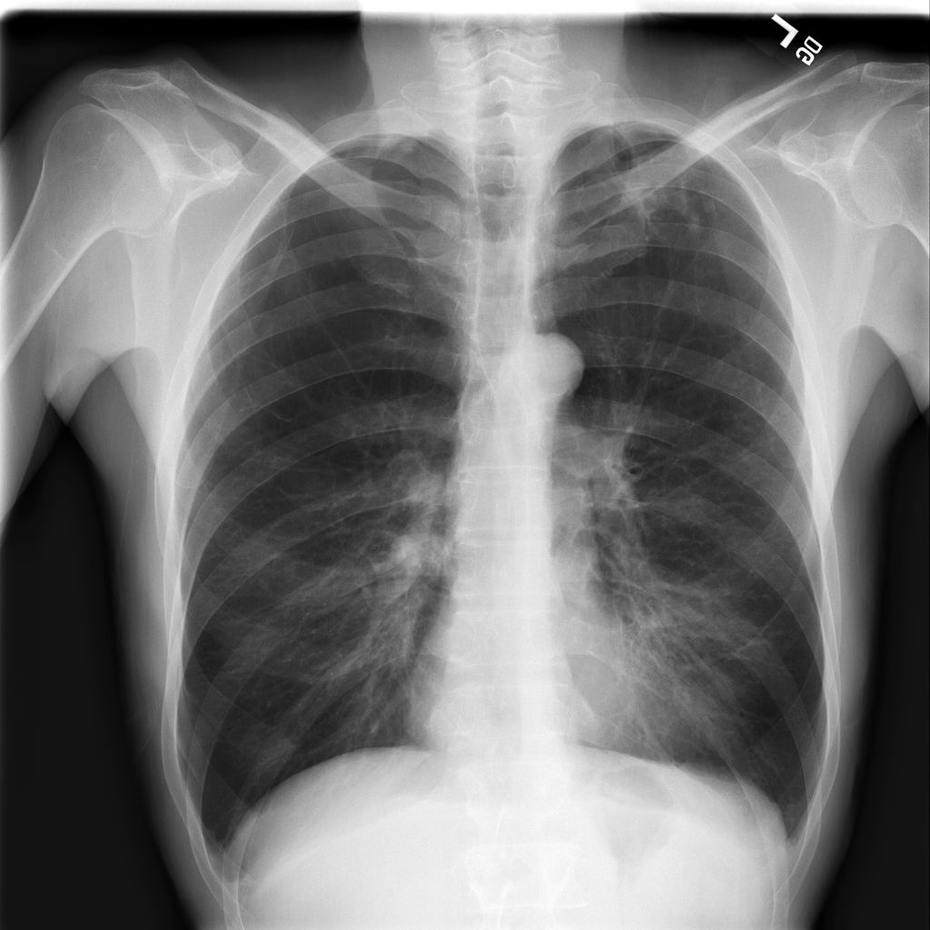
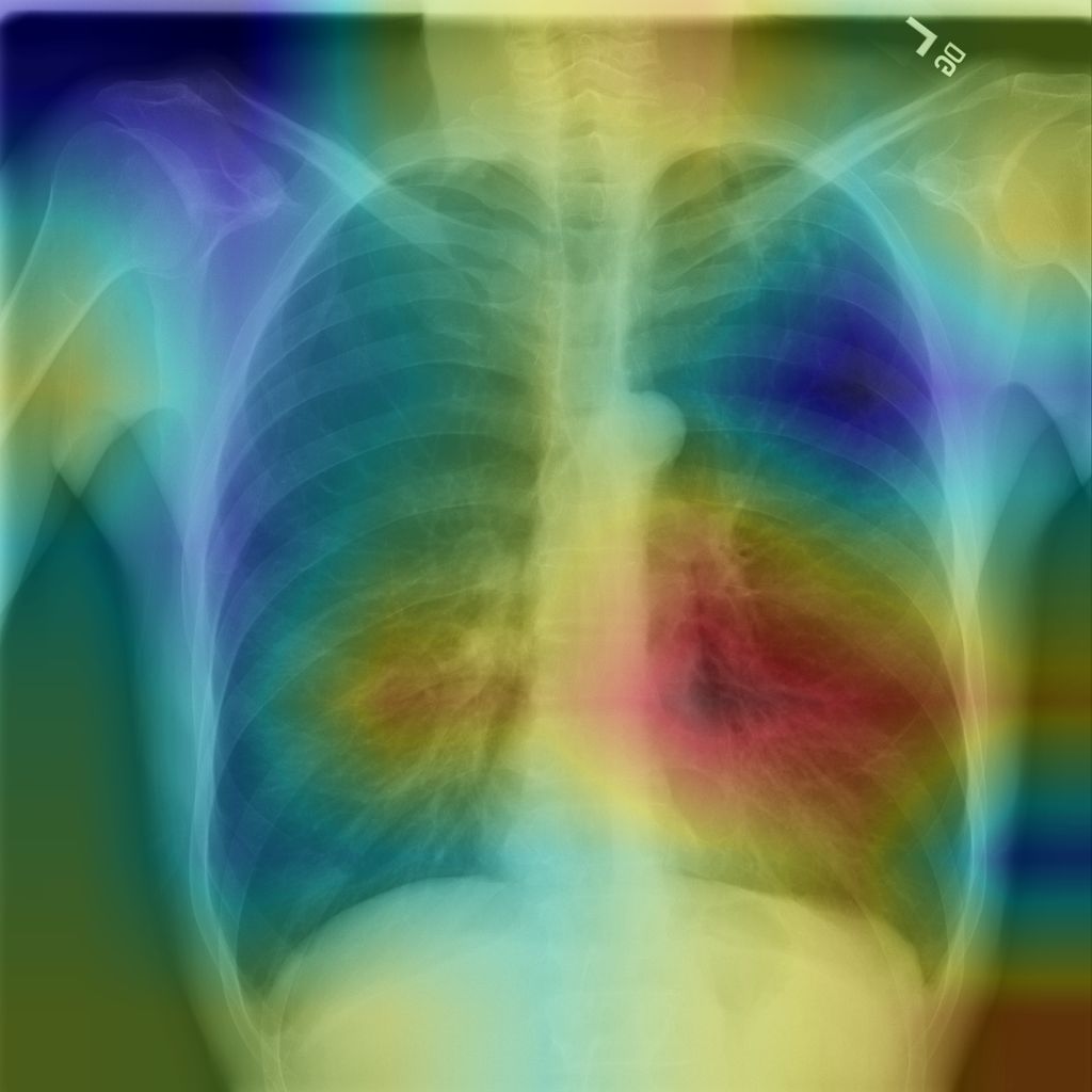
CheXNet is a 121-layer convolutional neural net-
work
that
takes
a
chest
X-ray
image
as
input,
and
outputs
the
probability
of
a
pathology.
On
this
example,
CheXnet
correctly
detects
pneumonia
and
also
localizes
areas
in
the
image
most
indicative
of
the
pathology.
Our
model,
ChexNet
(shown
in
Figure
1
),
is
a
121-
layer
convolutional
neural
network
that
inputs
a
chest
X-ray image and outputs the probability of pneumonia
along
with
a
heatmap
localizing
the
areas
of
the
im-
age
most
indicative
of
pneumonia.
We
train
CheXNet
on
the
recently
released
ChestX-ray14
dataset
(
Wang
et al.
,
2017
), which contains 112,120 frontal-view chest
X-ray images individually labeled with up to 14 differ-
ent
thoracic
diseases,
including
pneumonia.
We
use
arXiv:1711.05225v3 [cs.CV] 25 Dec 2017
CheXNet:
Radiologist-Level
Pneumonia
Detection
on
Chest
X-Rays
with
Deep
Learning
F1
Score
(95%
CI)
Radiologist
1
0.383
(0.309,
0.453)
Radiologist
2
0.356
(0.282,
0.428)
Radiologist
3
0.365
(0.291,
0.435)
Radiologist
4
0.442
(0.390,
0.492)
Radiologist
Avg.
0.387
(0.330,
0.442)
CheXNet
0.435
(0.387,
0.481)
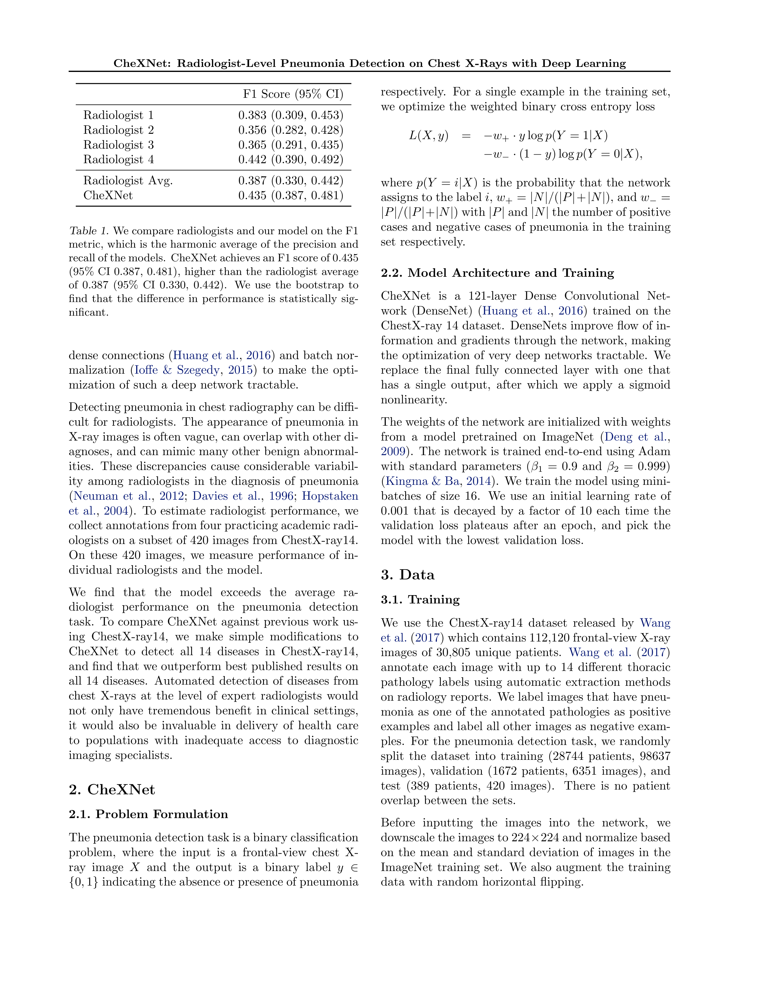
Table
1.
We compare radiologists and our model on the F1
metric,
which
is
the
harmonic
average
of
the
precision
and
recall of the models.
CheXNet achieves an F1 score of 0.435
(95%
CI
0.387,
0.481),
higher
than
the
radiologist
average
of
0.387
(95%
CI
0.330,
0.442).
We
use
the
bootstrap
to
find
that
the
difference
in
performance
is
statistically
sig-
nificant.
dense
connections
(
Huang
et
al.
,
2016
)
and
batch
nor-
malization
(
Ioffe
&
Szegedy
,
2015
)
to
make
the
opti-
mization
of
such
a
deep
network
tractable.
Detecting pneumonia in chest radiography can be diffi-
cult
for
radiologists.
The
appearance
of
pneumonia
in
X-ray images is often vague, can overlap with other di-
agnoses,
and
can
mimic
many
other
benign
abnormal-
ities.
These
discrepancies
cause
considerable
variabil-
ity
among
radiologists
in
the
diagnosis
of
pneumonia
(
Neuman
et
al.
,
2012
;
Davies
et
al.
,
1996
;
Hopstaken
et
al.
,
2004
).
To
estimate
radiologist
performance,
we
collect annotations from four practicing academic radi-
ologists
on
a
subset
of
420
images
from
ChestX-ray14.
On
these
420
images,
we
measure
performance
of
in-
dividual
radiologists
and
the
model.
We
find
that
the
model
exceeds
the
average
ra-
diologist
performance
on
the
pneumonia
detection
task.
To
compare
CheXNet
against
previous
work
us-
ing
ChestX-ray14,
we
make
simple
modifications
to
CheXNet
to
detect
all
14
diseases
in
ChestX-ray14,
and
find
that
we
outperform
best
published
results
on
all
14
diseases.
Automated
detection
of
diseases
from
chest
X-rays
at
the
level
of
expert
radiologists
would
not
only
have
tremendous
benefit
in
clinical
settings,
it
would
also
be
invaluable
in
delivery
of
health
care
to
populations
with
inadequate
access
to
diagnostic
imaging
specialists.
2.
CheXNet
2.1.
Problem
Formulation
The pneumonia detection task is a binary classification
problem,
where
the
input
is
a
frontal-view
chest
X-
ray
image
X
and
the
output
is
a
binary
label
y
∈
{
0
,
1
}
indicating the absence or presence of pneumonia
respectively.
For
a
single
example
in
the
training
set,
we
optimize
the
weighted
binary
cross
entropy
loss
L
(
X, y
)
=
−
w
+
·
y
log
p
(
Y
= 1
|
X
)
−
w
−
·
(1
−
y
) log
p
(
Y
= 0
|
X
)
,
where
p
(
Y
=
i
|
X
)
is
the
probability
that
the
network
assigns to the label
i
,
w
+
=
|
N
|
/
(
|
P
|
+
|
N
|
), and
w
−
=
|
P
|
/
(
|
P
|
+
|
N
|
) with
|
P
|
and
|
N
|
the number of positive
cases
and
negative
cases
of
pneumonia
in
the
training
set
respectively.
2.2.
Model
Architecture
and
Training
CheXNet
is
a
121-layer
Dense
Convolutional
Net-
work
(DenseNet)
(
Huang
et
al.
,
2016
)
trained
on
the
ChestX-ray
14
dataset.
DenseNets
improve
flow
of
in-
formation
and
gradients
through
the
network,
making
the
optimization
of
very
deep
networks
tractable.
We
replace
the
final
fully
connected
layer
with
one
that
has
a
single
output,
after
which
we
apply
a
sigmoid
nonlinearity.
The weights of the network are initialized with weights
from
a
model
pretrained
on
ImageNet
(
Deng
et
al.
,
2009
).
The
network
is
trained
end-to-end
using
Adam
with
standard
parameters
(
β
1
=
0
.
9
and
β
2
=
0
.
999)
(
Kingma & Ba
,
2014
).
We train the model using mini-
batches
of
size
16.
We
use
an
initial
learning
rate
of
0
.
001
that
is
decayed
by
a
factor
of
10
each
time
the
validation
loss
plateaus
after
an
epoch,
and
pick
the
model
with
the
lowest
validation
loss.
3.
Data
3.1.
Training
We
use
the
ChestX-ray14
dataset
released
by
Wang
et al.
(
2017
) which contains 112,120 frontal-view X-ray
images
of
30,805
unique
patients.
Wang
et
al.
(
2017
)
annotate
each
image
with
up
to
14
different
thoracic
pathology
labels
using
automatic
extraction
methods
on radiology reports.
We label images that have pneu-
monia
as
one
of
the
annotated
pathologies
as
positive
examples and label all other images as negative exam-
ples.
For
the
pneumonia
detection
task,
we
randomly
split
the
dataset
into
training
(28744
patients,
98637
images),
validation
(1672
patients,
6351
images),
and
test
(389
patients,
420
images).
There
is
no
patient
overlap
between
the
sets.
Before
inputting
the
images
into
the
network,
we
downscale the images to 224
×
224 and normalize based
on
the
mean
and
standard
deviation
of
images
in
the
ImageNet
training
set.
We
also
augment
the
training
data
with
random
horizontal
flipping.
CheXNet:
Radiologist-Level
Pneumonia
Detection
on
Chest
X-Rays
with
Deep
Learning
Pathology
Wang
et
al.
(2017)
Yao
et
al.
(2017)
CheXNet
(ours)
Atelectasis
0.716
0.772
0.8094
Cardiomegaly
0.807
0.904
0.9248
Effusion
0.784
0.859
0.8638
Infiltration
0.609
0.695
0.7345
Mass
0.706
0.792
0.8676
Nodule
0.671
0.717
0.7802
Pneumonia
0.633
0.713
0.7680
Pneumothorax
0.806
0.841
0.8887
Consolidation
0.708
0.788
0.7901
Edema
0.835
0.882
0.8878
Emphysema
0.815
0.829
0.9371
Fibrosis
0.769
0.767
0.8047
Pleural
Thickening
0.708
0.765
0.8062
Hernia
0.767
0.914
0.9164
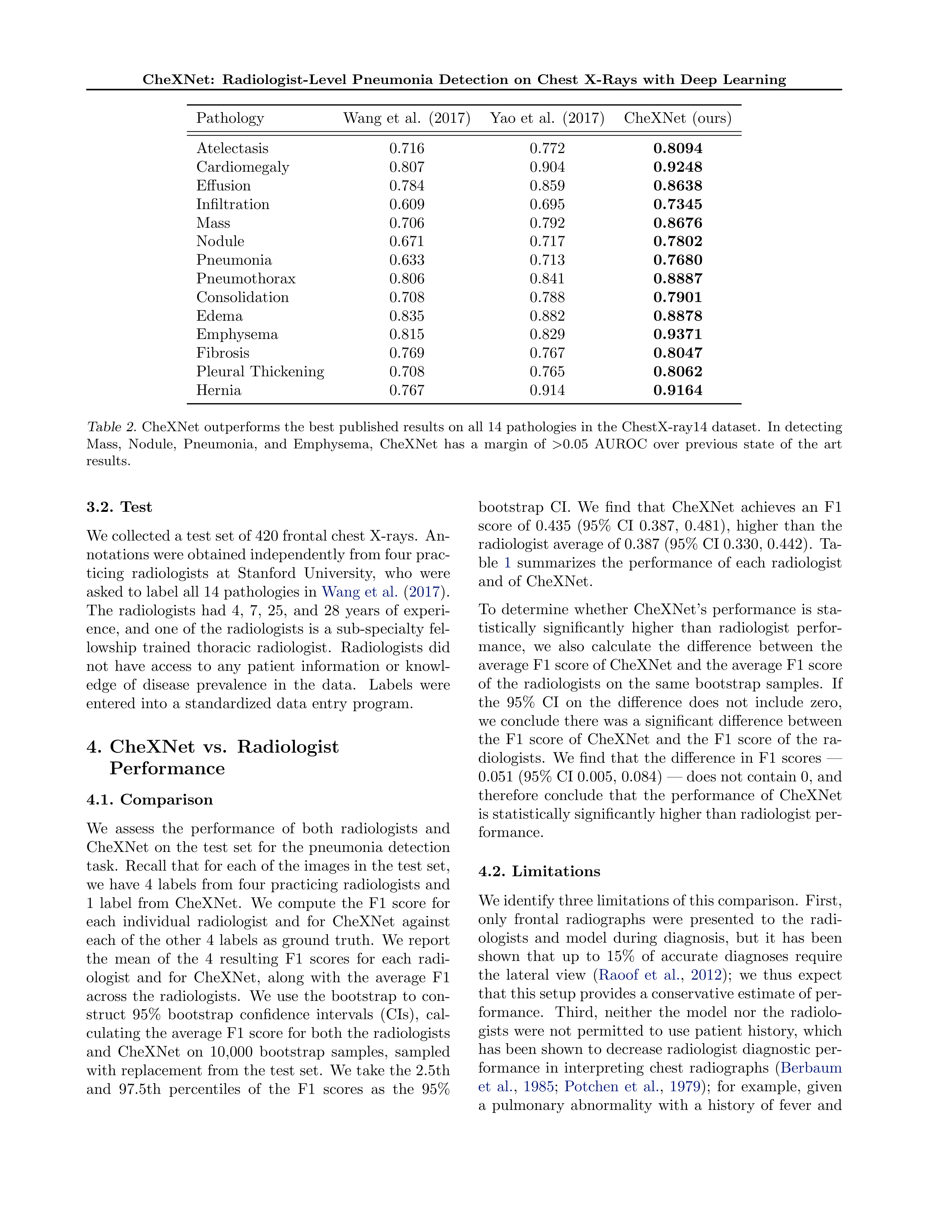
Table
2.
CheXNet
outperforms
the
best
published
results
on
all
14
pathologies
in
the
ChestX-ray14
dataset.
In
detecting
Mass,
Nodule,
Pneumonia,
and
Emphysema,
CheXNet
has
a
margin
of
>
0.05
AUROC
over
previous
state
of
the
art
results.
3.2.
Test
We collected a test set of 420 frontal chest X-rays.
An-
notations were obtained independently from four prac-
ticing
radiologists
at
Stanford
University,
who
were
asked to label all 14 pathologies in
Wang et al.
(
2017
).
The
radiologists
had
4,
7,
25,
and
28
years
of
experi-
ence,
and
one
of
the
radiologists
is
a
sub-specialty
fel-
lowship
trained
thoracic
radiologist.
Radiologists
did
not
have
access
to
any
patient
information
or
knowl-
edge
of
disease
prevalence
in
the
data.
Labels
were
entered
into
a
standardized
data
entry
program.
4.
CheXNet
vs.
Radiologist
Performance
4.1.
Comparison
We
assess
the
performance
of
both
radiologists
and
CheXNet
on
the
test
set
for
the
pneumonia
detection
task.
Recall that for each of the images in the test set,
we
have
4
labels
from
four
practicing
radiologists
and
1
label
from
CheXNet.
We
compute
the
F1
score
for
each
individual
radiologist
and
for
CheXNet
against
each
of
the
other
4
labels
as
ground
truth.
We
report
the
mean
of
the
4
resulting
F1
scores
for
each
radi-
ologist
and
for
CheXNet,
along
with
the
average
F1
across
the
radiologists.
We
use
the
bootstrap
to
con-
struct
95%
bootstrap
confidence
intervals
(CIs),
cal-
culating
the
average
F1
score
for
both
the
radiologists
and
CheXNet
on
10,000
bootstrap
samples,
sampled
with
replacement
from
the
test
set.
We
take
the
2.5th
and
97.5th
percentiles
of
the
F1
scores
as
the
95%
bootstrap
CI.
We
find
that
CheXNet
achieves
an
F1
score
of
0.435
(95%
CI
0.387,
0.481),
higher
than
the
radiologist average of 0.387 (95% CI 0.330, 0.442).
Ta-
ble
1
summarizes
the
performance
of
each
radiologist
and
of
CheXNet.
To
determine
whether
CheXNet’s
performance
is
sta-
tistically
significantly
higher
than
radiologist
perfor-
mance,
we
also
calculate
the
difference
between
the
average F1 score of CheXNet and the average F1 score
of
the
radiologists
on
the
same
bootstrap
samples.
If
the
95%
CI
on
the
difference
does
not
include
zero,
we
conclude
there
was
a
significant
difference
between
the
F1
score
of
CheXNet
and
the
F1
score
of
the
ra-
diologists.
We
find
that
the
difference
in
F1
scores
—
0.051 (95% CI 0.005, 0.084) — does not contain 0, and
therefore
conclude
that
the
performance
of
CheXNet
is statistically significantly higher than radiologist per-
formance.
4.2.
Limitations
We identify three limitations of this comparison.
First,
only
frontal
radiographs
were
presented
to
the
radi-
ologists
and
model
during
diagnosis,
but
it
has
been
shown
that
up
to
15%
of
accurate
diagnoses
require
the
lateral
view
(
Raoof
et
al.
,
2012
);
we
thus
expect
that this setup provides a conservative estimate of per-
formance.
Third,
neither
the
model
nor
the
radiolo-
gists
were
not
permitted
to
use
patient
history,
which
has been shown to decrease radiologist diagnostic per-
formance
in
interpreting
chest
radiographs
(
Berbaum
et
al.
,
1985
;
Potchen
et
al.
,
1979
);
for
example,
given
a
pulmonary
abnormality
with
a
history
of
fever
and
CheXNet:
Radiologist-Level
Pneumonia
Detection
on
Chest
X-Rays
with
Deep
Learning
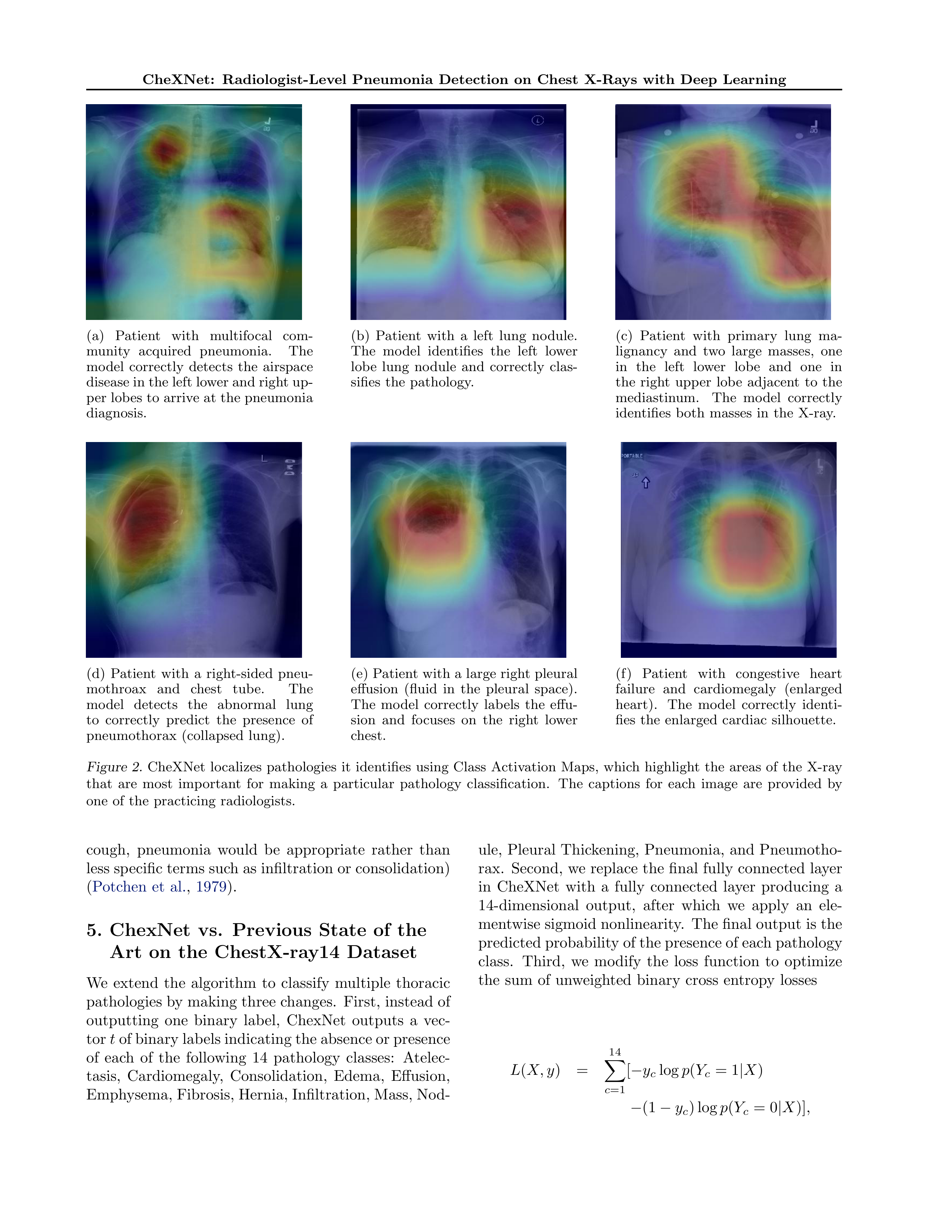
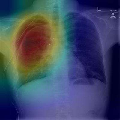
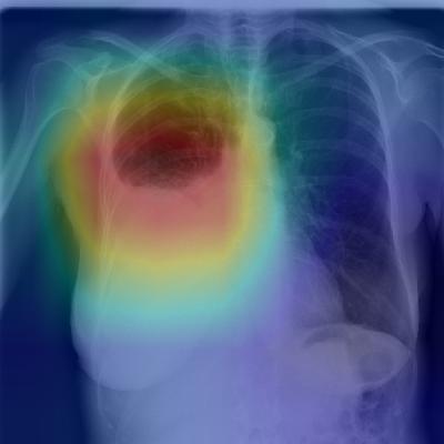
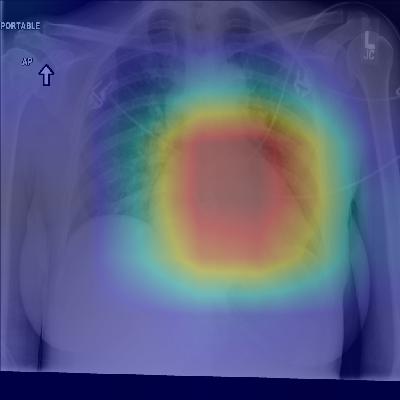
(a)
Patient
with
multifocal
com-
munity
acquired
pneumonia.
The
model
correctly
detects
the
airspace
disease in the left lower and right up-
per lobes to arrive
at
the
pneumonia
diagnosis.
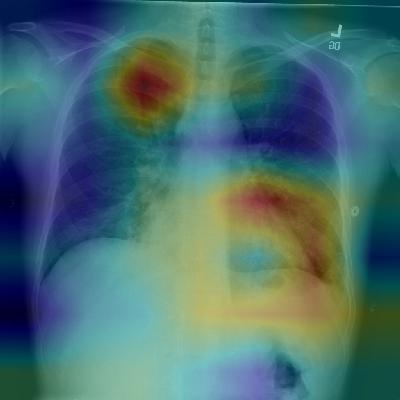
(b)
Patient
with
a
left
lung
nodule.
The
model
identifies
the
left
lower
lobe
lung
nodule
and
correctly
clas-
sifies
the
pathology.
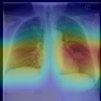
(c)
Patient
with
primary
lung
ma-
lignancy
and
two
large
masses,
one
in
the
left
lower
lobe
and
one
in
the
right
upper
lobe
adjacent
to
the
mediastinum.
The
model
correctly
identifies
both
masses
in
the
X-ray.
(d)
Patient
with
a
right-sided
pneu-
mothroax
and
chest
tube.
The
model
detects
the
abnormal
lung
to
correctly
predict
the
presence
of
pneumothorax
(collapsed
lung).
(e)
Patient
with
a
large
right
pleural
effusion
(fluid
in
the
pleural
space).
The
model
correctly
labels
the
effu-
sion
and
focuses
on
the
right
lower
chest.
(f)
Patient
with
congestive
heart
failure
and
cardiomegaly
(enlarged
heart).
The
model
correctly
identi-
fies
the
enlarged
cardiac
silhouette.
Figure
2.
CheXNet
localizes
pathologies
it
identifies
using
Class
Activation
Maps,
which
highlight
the
areas
of
the
X-ray
that
are
most
important
for
making
a
particular
pathology
classification.
The
captions
for
each
image
are
provided
by
one
of
the
practicing
radiologists.
cough,
pneumonia
would
be
appropriate
rather
than
less specific terms such as infiltration or consolidation)
(
Potchen
et
al.
,
1979
).
5.
ChexNet
vs.
Previous
State
of
the
Art
on
the
ChestX-ray14
Dataset
We
extend
the
algorithm
to
classify
multiple
thoracic
pathologies by making three changes.
First, instead of
outputting
one
binary
label,
ChexNet
outputs
a
vec-
tor
t
of binary labels indicating the absence or presence
of
each
of
the
following
14
pathology
classes:
Atelec-
tasis,
Cardiomegaly,
Consolidation,
Edema,
Effusion,
Emphysema, Fibrosis, Hernia, Infiltration, Mass, Nod-
ule,
Pleural
Thickening,
Pneumonia,
and
Pneumotho-
rax.
Second,
we
replace
the
final
fully
connected
layer
in
CheXNet
with
a
fully
connected
layer
producing
a
14-dimensional
output,
after
which
we
apply
an
ele-
mentwise sigmoid nonlinearity.
The final output is the
predicted probability of the presence of each pathology
class.
Third,
we
modify
the
loss
function
to
optimize
the
sum
of
unweighted
binary
cross
entropy
losses
L
(
X, y
)
=
14
X
c
=1
[
−
y
c
log
p
(
Y
c
= 1
|
X
)
−
(1
−
y
c
) log
p
(
Y
c
= 0
|
X
)]
,
CheXNet:
Radiologist-Level
Pneumonia
Detection
on
Chest
X-Rays
with
Deep
Learning
where
p
(
Y
c
=
1
|
X
)
is
the
predicted
probability
that
the
image
contains
the
pathology
c
and
p
(
Y
c
=
0
|
X
)
is
the
predicted
probability
that
the
image
does
not
contain
the
pathology
c
.
We randomly split the dataset into training (70%), val-
idation
(10%),
and
test
(20%)
sets,
following
previous
work
on
ChestX-ray14
(
Wang
et
al.
,
2017
;
Yao
et
al.
,
2017
).
We
ensure
that
there
is
no
patient
overlap
be-
tween the splits.
We compare the per-class AUROC of
the model against the previous state of the art held by
Yao
et
al.
(
2017
)
on
13
classes
and
Wang
et
al.
(
2017
)
on
the
remaining
1
class.
We find that CheXNet achieves state of the art results
on all 14 pathology classes.
Table
2
illustrates the per-
class
AUROC
comparison
on
the
test
set.
On
Mass,
Nodule,
Pneumonia,
and
Emphysema,
we
outperform
previous
state
of
the
art
considerably
(
>
0
.
05
increase
in
AUROC).
6.
Model
Interpretation
To
interpret
the
network
predictions,
we
also
produce
heatmaps
to
visualize
the
areas
of
the
image
most
in-
dicative
of
the
disease
using
class
activation
mappings
(CAMs)
(
Zhou
et
al.
,
2016
).
To
generate
the
CAMs,
we
feed
an
image
into
the
fully
trained
network
and
extract
the
feature
maps
that
are
output
by
the
final
convolutional layer.
Let
f
k
be the
k
th feature map and
let
w
c,k
be
the
weight
in
the
final
classification
layer
for
feature
map
k
leading
to
pathology
c
.
We
obtain
a
map
M
c
of
the
most
salient
features
used
in
classi-
fying
the
image
as
having
pathology
c
by
taking
the
weighted
sum
of
the
feature
maps
using
their
associ-
ated
weights.
Formally,
M
c
=
X
k
w
c,k
f
k
.
We
identify
the
most
important
features
used
by
the
model
in
its
prediction
of
the
pathology
c
by
upscal-
ing
the
map
M
c
to
the
dimensions
of
the
image
and
overlaying
the
image.
Figure
2
shows several examples of CAMs on the pneu-
monia
detection
task
as
well
as
the
14-class
pathology
classification
task.
7.
Related
Work
Recent
advancements
in
deep
learning
and
large
datasets
have
enabled
algorithms
to
surpass
the
per-
formance
of
medical
professionals
in
a
wide
variety
of
medical
imaging
tasks,
including
diabetic
retinopathy
detection (
Gulshan et al.
,
2016
), skin cancer classifica-
tion
(
Esteva
et
al.
,
2017
),
arrhythmia
detection
(
Ra-
jpurkar
et
al.
,
2017
),
and
hemorrhage
identification
(
Grewal
et
al.
,
2017
).
Automated
diagnosis
from
chest
radiographs
has
re-
ceived
increasing
attention
with
algorithms
for
pul-
monary
tuberculosis
classification
(
Lakhani
&
Sun-
daram
,
2017
) and lung nodule detection (
Huang et al.
,
2017
).
Islam
et
al.
(
2017
)
studied
the
performance
of
various
convolutional
architectures
on
different
ab-
normalities
using
the
publicly
available
OpenI
dataset
(
Demner-Fushman
et
al.
,
2015
).
Wang
et
al.
(
2017
)
released
ChestX-ray-14,
an
order
of
magnitude
larger
than
previous
datasets
of
its
kind,
and
also
bench-
marked
different
convolutional
neural
network
archi-
tectures
pre-trained
on
ImageNet.
Recently
Yao
et
al.
(
2017
)
exploited
statistical
dependencies
between
la-
bels
in
order
make
more
accurate
predictions,
outper-
forming
Wang
et
al.
(
2017
)
on
13
of
14
classes.
8.
Conclusion
Pneumonia
accounts
for
a
significant
proportion
of
patient
morbidity
and
mortality
(
Gon¸calves-Pereira
et
al.
,
2013
).
Early
diagnosis
and
treatment
of
pneu-
monia is critical to preventing complications including
death
(
Aydogdu
et
al.
,
2010
).
With
approximately
2
billion
procedures
per
year,
chest
X-rays
are
the
most
common
imaging
examination
tool
used
in
practice,
critical
for
screening,
diagnosis,
and
management
of
a
variety
of
diseases
including
pneumonia
(
Raoof
et
al.
,
2012
).
However,
two
thirds
of
the
global
population
lacks
access
to
radiology
diagnostics,
according
to
an
estimate
by
the
World
Health
Organization
(
Mollura
et al.
,
2010
).
There is a shortage of experts who can in-
terpret X-rays,
even when imaging equipment is avail-
able, leading to increased mortality from treatable dis-
eases
(
Kesselman
et
al.
,
2016
).
We
develop
an
algorithm
which
detects
pneumonia
from
frontal-view
chest
X-ray
images
at
a
level
ex-
ceeding
practicing
radiologists.
We
also
show
that
a
simple
extension
of
our
algorithm
to
detect
multi-
ple
diseases
outperforms
previous
state
of
the
art
on
ChestX-ray14,
the
largest
publicly
available
chest
X-
ray
dataset.
With
automation
at
the
level
of
experts,
we
hope
that
this
technology
can
improve
healthcare
delivery
and
increase
access
to
medical
imaging
ex-
pertise
in
parts
of
the
world
where
access
to
skilled
radiologists
is
limited.
CheXNet:
Radiologist-Level
Pneumonia
Detection
on
Chest
X-Rays
with
Deep
Learning
9.
Acknowledgements
We
would
like
to
acknowledge
the
Stanford
Center
for
Artificial Intelligence in Medicine and Imaging for clin-
ical
dataset
infrastructure
support
(
AIMI.stanford.
edu
).
References
Aydogdu,
M,
Ozyilmaz,
E,
Aksoy,
Handan,
Gursel,
G,
and
Ekim,
Numan.
Mortality
prediction
in
community-acquired
pneumonia
requiring
mechan-
ical
ventilation;
values
of
pneumonia
and
intensive
care
unit
severity
scores.
Tuberk
Toraks
,
58(1):25–
34,
2010.
Berbaum,
K,
Franken
Jr,
EA,
and
Smith,
WL.
The
effect
of
comparison
films
upon
resident
interpre-
tation
of
pediatric
chest
radiographs.
Investigative
radiology
,
20(2):124–128,
1985.
CDC,
2017.
URL
https://www.cdc.gov/features/
pneumonia/index.html
.
Cherian, Thomas, Mulholland, E Kim, Carlin, John B,
Ostensen,
Harald,
Amin,
Ruhul,
Campo,
Mar-
garet
de,
Greenberg,
David,
Lagos,
Rosanna,
Lucero,
Marilla,
Madhi,
Shabir
A,
et
al.
Standard-
ized
interpretation
of
paediatric
chest
radiographs
for
the
diagnosis
of
pneumonia
in
epidemiological
studies.
Bulletin
of
the
World
Health
Organization
,
83(5):353–359,
2005.
Davies,
H
Dele,
Wang,
Elaine
E-l,
Manson,
David,
Babyn,
Paul,
and
Shuckett,
Bruce.
Reliability
of
the
chest
radiograph
in
the
diagnosis
of
lower
res-
piratory
infections
in
young
children.
The
Pediatric
infectious
disease
journal
,
15(7):600–604,
1996.
Demner-Fushman,
Dina,
Kohli,
Marc
D,
Rosenman,
Marc
B,
Shooshan,
Sonya
E,
Rodriguez,
Laritza,
Antani,
Sameer,
Thoma,
George
R,
and
McDonald,
Clement
J.
Preparing
a
collection
of
radiology
ex-
aminations for distribution and retrieval.
Journal
of
the
American
Medical
Informatics
Association
,
23
(2):304–310,
2015.
Deng,
Jia,
Dong,
Wei,
Socher,
Richard,
Li,
Li-Jia,
Li,
Kai,
and
Fei-Fei,
Li.
Imagenet:
A
large-scale
hier-
archical
image
database.
In
Computer
Vision
and
Pattern Recognition,
2009. CVPR 2009. IEEE Con-
ference
on
,
pp.
248–255.
IEEE,
2009.
Esteva,
Andre,
Kuprel,
Brett,
Novoa,
Roberto
A,
Ko,
Justin,
Swetter,
Susan
M,
Blau,
Helen
M,
and
Thrun,
Sebastian.
Dermatologist-level
classification
of
skin
cancer
with
deep
neural
networks.
Nature
,
542(7639):115–118,
2017.
Franquet,
T.
Imaging
of
pneumonia:
trends
and
algo-
rithms.
European
Respiratory
Journal
,
18(1):196–
208,
2001.
Gon¸calves-Pereira,
Jo˜ao,
Concei¸c˜ao,
Catarina,
and
P´ovoa,
Pedro.
Community-acquired
pneumo-
nia:
identification
and
evaluation
of
nonresponders.
Therapeutic
advances
in
infectious
disease
,
1(1):5–
17,
2013.
Grewal,
Monika,
Srivastava,
Muktabh
Mayank,
Ku-
mar,
Pulkit,
and
Varadarajan,
Srikrishna.
Radnet:
Radiologist
level
accuracy
using
deep
learning
for
hemorrhage
detection
in
ct
scans.
arXiv
preprint
arXiv:1710.04934
,
2017.
Gulshan,
Varun,
Peng,
Lily,
Coram,
Marc,
Stumpe,
Martin
C,
Wu,
Derek,
Narayanaswamy,
Arunacha-
lam,
Venugopalan,
Subhashini,
Widner,
Kasumi,
Madams,
Tom,
Cuadros,
Jorge,
et
al.
Development
and
validation
of
a
deep
learning
algorithm
for
de-
tection of diabetic retinopathy in retinal fundus pho-
tographs.
Jama
,
316(22):2402–2410,
2016.
Hopstaken,
RM,
Witbraad,
T,
Van
Engelshoven,
JMA,
and
Dinant,
GJ.
Inter-observer
variation
in
the
interpretation
of
chest
radiographs
for
pneumo-
nia
in
community-acquired
lower
respiratory
tract
infections.
Clinical
radiology
,
59(8):743–752,
2004.
Huang,
Gao,
Liu,
Zhuang,
Weinberger,
Kilian
Q,
and
van der Maaten, Laurens.
Densely connected convo-
lutional networks.
arXiv preprint arXiv:1608.06993
,
2016.
Huang,
Peng,
Park,
Seyoun,
Yan,
Rongkai,
Lee,
Junghoon,
Chu,
Linda
C,
Lin,
Cheng
T,
Hussien,
Amira,
Rathmell,
Joshua,
Thomas,
Brett,
Chen,
Chen, et al. Added value of computer-aided ct image
features
for
early
lung
cancer
diagnosis
with
small
pulmonary
nodules:
A
matched
case-control
study.
Radiology
,
pp.
162725,
2017.
Ioffe, Sergey and Szegedy, Christian.
Batch normaliza-
tion:
Accelerating
deep
network
training
by
reduc-
ing internal covariate shift.
In
International
Confer-
ence
on
Machine
Learning
,
pp.
448–456,
2015.
Islam,
Mohammad
Tariqul,
Aowal,
Md
Abdul,
Min-
haz,
Ahmed
Tahseen,
and
Ashraf,
Khalid.
Ab-
normality
detection
and
localization
in
chest
x-rays
using
deep
convolutional
neural
networks.
arXiv
preprint
arXiv:1705.09850
,
2017.
Kesselman,
Andrew,
Soroosh,
Garshasb,
Mollura,
Daniel
J,
and
Group,
RAD-AID
Conference
Writ-
ing.
2015
rad-aid
conference
on
international
radi-
ology
for
developing
countries:
The
evolving
global
CheXNet:
Radiologist-Level
Pneumonia
Detection
on
Chest
X-Rays
with
Deep
Learning
radiology
landscape.
Journal
of
the
American
Col-
lege
of
Radiology
,
13(9):1139–1144,
2016.
Kingma,
Diederik
and
Ba,
Jimmy.
Adam:
A
method
for
stochastic
optimization.
arXiv
preprint
arXiv:1412.6980
,
2014.
Lakhani,
Paras and Sundaram,
Baskaran.
Deep learn-
ing
at
chest
radiography:
Automated
classification
of
pulmonary
tuberculosis
by
using
convolutional
neural
networks.
Radiology
,
pp.
162326,
2017.
Mollura,
Daniel
J,
Azene,
Ezana
M,
Starikovsky,
Anna,
Thelwell,
Aduke,
Iosifescu,
Sarah,
Kimble,
Cary,
Polin,
Ann,
Garra,
Brian
S,
DeStigter,
Kris-
ten
K,
Short,
Brad,
et
al.
White
paper
report
of
the rad-aid conference on international radiology for
developing
countries:
identifying
challenges,
oppor-
tunities,
and
strategies
for
imaging
services
in
the
developing
world.
Journal
of
the
American
College
of
Radiology
,
7(7):495–500,
2010.
Neuman,
Mark
I,
Lee,
Edward
Y,
Bixby,
Sarah,
Diperna,
Stephanie,
Hellinger,
Jeffrey,
Markowitz,
Richard,
Servaes,
Sabah,
Monuteaux,
Michael
C,
and Shah,
Samir S.
Variability in the interpretation
of
chest
radiographs
for
the
diagnosis
of
pneumo-
nia
in
children.
Journal
of
hospital
medicine
,
7(4):
294–298,
2012.
Potchen,
EJ,
Gard,
JW,
Lazar,
P,
Lahaie,
P,
and
Andary,
M.
Effect
of
clinical
history
data
on
chest
film interpretation-direction or distraction. In
Inves-
tigative
Radiology
,
volume
14,
pp.
404–404,
1979.
Rajpurkar,
Pranav,
Hannun,
Awni
Y,
Haghpanahi,
Masoumeh,
Bourn,
Codie,
and
Ng,
Andrew
Y.
Cardiologist-level
arrhythmia
detection
with
con-
volutional
neural
networks.
arXiv
preprint
arXiv:1707.01836
,
2017.
Raoof,
Suhail,
Feigin,
David,
Sung,
Arthur,
Raoof,
Sabiha,
Irugulpati,
Lavanya,
and
Rosenow,
Edward
C.
Interpretation
of
plain
chest
roentgenogram.
CHEST
Journal
,
141(2):545–558,
2012.
Wang,
Xiaosong,
Peng,
Yifan,
Lu,
Le,
Lu,
Zhiyong,
Bagheri, Mohammadhadi, and Summers, Ronald M.
Chestx-ray8:
Hospital-scale
chest
x-ray
database
and
benchmarks
on
weakly-supervised
classification
and
localization
of
common
thorax
diseases.
arXiv
preprint
arXiv:1705.02315
,
2017.
WHO.
Standardization
of
interpretation
of
chest
ra-
diographs
for
the
diagnosis
of
pneumonia
in
chil-
dren.
2001.
Yao,
Li,
Poblenz,
Eric,
Dagunts,
Dmitry,
Covington,
Ben,
Bernard,
Devon,
and
Lyman,
Kevin.
Learning
to
diagnose
from
scratch
by
exploiting
dependen-
cies among labels.
arXiv preprint arXiv:1710.10501
,
2017.
Zhou,
Bolei,
Khosla,
Aditya,
Lapedriza,
Agata,
Oliva,
Aude,
and
Torralba,
Antonio.
Learning
deep
fea-
tures
for
discriminative
localization.
In
Proceedings
of
the
IEEE
Conference
on
Computer
Vision
and
Pattern
Recognition
,
pp.
2921–2929,
2016.